
NewsMakers
Osteoporosis could prove deadly in postmenopausal women
A new study suggests it may also increase a woman’s overall risk of death—by as much as 47%—especially within specific ranges of bone mineral density (0.46-0.71 g/cm2 for total femur bone mineral density).


Osteoporosis, which is highly prevalent in postmenopausal women, has long been associated with an increased risk of fractures. A new study suggests it may also increase a woman’s overall risk of death—by as much as 47%—especially within specific ranges of bone mineral density (0.46-0.71 g/cm2 for total femur bone mineral density).
Results of the study are published online in Menopause, the journal of The Menopause Society.
As the total population ages, the incidence of osteoporosis also increases. In 2022, the global prevalence of osteoporosis was 19.7%, with women exhibiting a significantly higher prevalence than men (23.1%). One study projected that by 2030 the number of people affected by osteoporosis worldwide will reach 263 million, with 154 million of them being women. Previous research has documented that postmenopausal women experience a significantly higher mortality rate within 1 year after hip or vertebral fractures.
The decline of estrogen levels during the menopause transition has been linked to a number of physiologic changes across multiple systems, including bone metabolism, cardiovascular function, muscle mass, and fat distribution. Regarding bone health, declining estrogen levels accelerate bone resorption and inhibit bone formation, leading to a rapid decrease in bone mineral density (especially in the femoral region), which in turn increases the risk of osteoporosis and fractures.
Most research to date has focused on the association between low bone mineral density and adverse outcomes, such as falls and fractures. However, there is a lack of systematic studies examining whether increasing bone density in postmenopausal women can reduce the risk of death. In this new study involving nearly 3,000 postmenopausal women, bone mineral density at four femoral sites was assessed using dual-energy x-ray absorptiometry.
The analysis revealed that mortality risk was significantly elevated when femoral bone mineral density reached the osteoporotic threshold or in the presence of osteoporotic fractures. After full adjustment, osteoporosis was associated with a 47% increased risk of mortality. A stronger inverse association between increased bone mineral density and mortality risk was observed within specific ranges, suggesting that bone mineral density should serve as a prognostic biomarker of systemic health.
Study results are published in the article “Femoral bone mineral density and mortality risk in postmenopausal women: a National Health and Nutrition Examination Survey cohort study.”
“Osteoporosis often remains a silent threat after menopause, despite its profound effect on women’s lives—from loss of height, poor balance, and reduced mobility to disfigurement, pain, and even premature death. Early screening and preventive measures, including a calcium-rich diet (preferably from food sources), regular weight-bearing exercise, and hormone therapy when appropriate, can significantly improve bone health and reduce risks not only of fractures but also cardiovascular disease, certain cancers, and dementia. It’s time we bring this conversation to the forefront,” says Dr. Monica Christmas, associate medical director for The Menopause Society.







NewsMakers
The look and feel of your neighborhood may affect your sleep
Those living in neighborhoods rated as having a stronger sense of safety tended to sleep longer, and that this rating appeared to be shaped by the streetscape.

Your neighborhood’s “streetscape”—the physical environment of your street—is something that you have probably never consciously thought about, despite seeing it every day. But what if the streetscape was affecting you on a subconscious level and disrupting your sleep?
This intriguing question is posed by the findings of a study led by Professor Daisuke Matsushita of the Graduate School of Human Life and Ecology at Osaka Metropolitan University. The research team used AI to analyze more than 200,000 Google Street View images to identify visual neighborhood characteristics. They then linked these characteristics to the self-reported sleep of 1,089 working adults living on the lower floors of apartments in Tokyo, who were most likely to be affected by the streetscape.
They found that those living in neighborhoods rated as having a stronger sense of safety tended to sleep longer, and that this rating appeared to be shaped by the streetscape. Generally, people slept longer in areas with lots of greenery, such as leafy trees, on the street. Similarly, high “enclosure”—meaning many tall vertical buildings and few wide-open spaces—was also associated with longer sleep and fewer insomnia symptoms.
However, the study also made a surprising finding. The researchers found that highly walkable streetscapes, such as those with more sidewalks and traffic signs, were associated with a lowered sense of safety and shortened sleep duration.
This suggests that walkability does not always represent a reassuring environment. Instead, a possible explanation is that streets with extensive pedestrian infrastructure are often busier, more crowded, and used by more strangers, which may be perceived as less safe or less relaxing than quieter residential streets.
“This study demonstrates the potential for evaluating streetscape characteristics across large geographic areas in a cost-effective manner,” Dr. Matsushita said. “Based on the technique used in this study, cities could potentially measure perceived safety, beauty, liveliness, and enclosure as well as pollution and noise.”
“We hope that opening up this new perspective creates further possibilities for designing healthier neighborhoods,” he concludes.
The findings were published in Building and Environment.
NewsMakers
Workout or nap? Either can help your sleepless brain, study finds
People who either completed 20 minutes of moderate-to-vigorous exercise or took a 90-minute nap performed about 22 per cent better on memory tests after 30 hours without sleep than those who did neither.

A short workout or a nap can help protect memory after a sleepless night, a McGill University-led study has found.
Researchers found people who either completed 20 minutes of moderate-to-vigorous exercise or took a 90-minute nap performed about 22 per cent better on memory tests after 30 hours without sleep than those who did neither. The findings, published in Proceedings of the National Academy of Sciences (PNAS), point to practical ways to counter the effects of sleep deprivation.
“Sleep loss affects nearly every aspect of how we think and function, but many people can’t simply stop what they’re doing and get more sleep,” said senior author Marc Roig, Professor in McGill’s School of Physical and Occupational Therapy. “Our findings show that even a brief bout of exercise may help preserve one of our most important cognitive abilities.”
The study involved 54 healthy young adults who stayed awake for 30 consecutive hours under lab supervision. Participants were then assigned to one of three groups: a 20-minute cycling session, a 90-minute nap or a control condition. Three days later, researchers tested their memory for images they had viewed immediately after the intervention. Those who exercised or napped remembered significantly more images than participants who did neither.
Same result, different pathways
While the memory benefits were nearly identical, brain recordings revealed that exercise and napping helped in different ways.
Napping appeared to help the brain recharge, making it easier to take in and remember new information. Exercise, by contrast, helped the brain use its remaining resources more efficiently, without making participants feel more tired.
The findings could eventually inform fatigue-management strategies in workplaces where sleep loss is common and mistakes can have serious consequences, such as health care, transportation and emergency response.
“A nap isn’t always possible in the middle of a shift,” said first author Madhura Lotlikar, a doctoral candidate in McGill’s Department of Neurology and Neurosurgery. “Exercise is accessible, inexpensive and easy to implement. That makes it a promising tool to help people stay cognitively sharp when sleep is limited.”
The researchers emphasize that exercise cannot replace sleep, but it may help people function better when getting enough rest isn’t possible.
About the study
“Protecting episodic memory after sleep loss: Similar benefits of exercise and naps via distinct neural contributions” by Madhura Lotlikar and Marc Roig et al., was published in Proceedings of the National Academy of Sciences of the United States of America.
NewsMakers
Study links coffee consumption to metabolic health and sex hormones
Despite having a similar body mass index (BMI), individuals with higher coffee consumption had lower total and visceral fat and greater skeletal muscle mass than those who consumed less coffee.
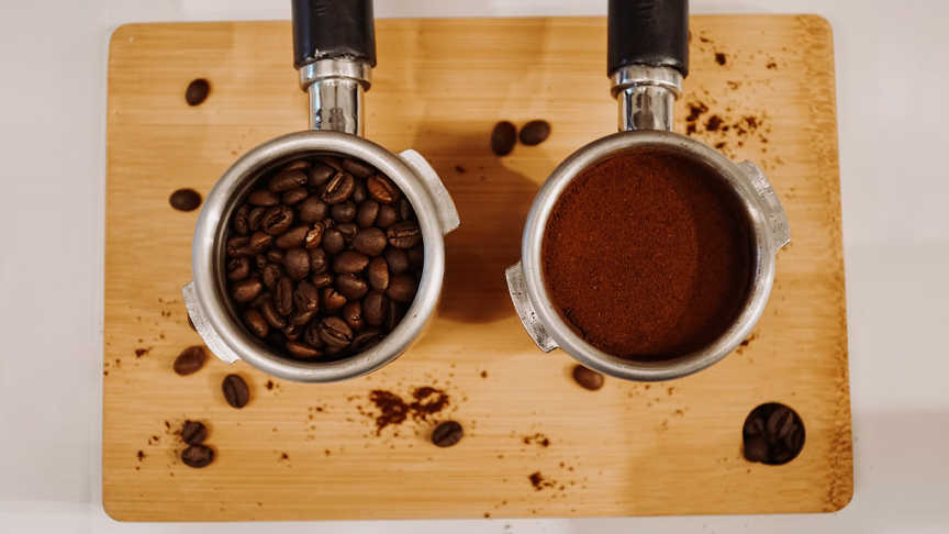
Coffee is one of the world’s most widely consumed beverages, and previous research has linked its consumption to a lower risk of conditions such as type 2 diabetes and cardiovascular disease. However, the biological mechanisms behind these benefits remain unclear. A new Finnish study links habitual coffee consumption to healthier body composition and metabolic markers, while revealing distinct associations with sex hormones in men and women.
The study, conducted at the University of Oulu, analysed data from 2,264 participants aged 46 in the Northern Finland Birth Cohort 1966. Researchers examined how habitual coffee consumption was associated with circulating metabolites, cardiometabolic risk markers and sex hormones.
Despite having a similar body mass index (BMI), individuals with higher coffee consumption had lower total and visceral fat and greater skeletal muscle mass than those who consumed less coffee.
In both men and women, higher coffee consumption was correlated with lower circulating levels of branched-chain amino acids, biomarkers that have previously been linked to insulin resistance and an increased risk of type 2 diabetes when chronically elevated.
The strongest sex-specific associations were observed in men. Higher coffee consumption was linked to a more favourable glucose–insulin profile, higher concentrations of total and bioavailable testosterone, and increased levels of sex hormone-binding globulin (SHBG). At the same time, free testosterone and the free androgen index were modestly lower. In women, hormonal associations were more limited and were primarily characterised by higher SHBG and lower measures of free androgens.
“Coffee is consumed by millions of people every day, yet we still know surprisingly little about how it relates to our metabolism and hormones. What stood out in our findings was a distinct hormonal signature that didn’t disappear even after we took into account BMI and lifestyle factors, with several of these associations differing between men and women,” says Luca Verroest, lead author of the study and Doctoral Researcher at the University of Oulu.
The results suggest that hormonal pathways may partly explain the relationship between coffee consumption and metabolic health. However, as this was an observational study, the findings demonstrate associations rather than cause-and-effect relationships.
The study is particularly relevant in Finland, one of the world’s highest coffee-consuming countries, where annual consumption averages around 11.8 kilograms per person.
The researchers say the findings provide a foundation for future studies aimed at determining whether coffee itself drives these biological changes and identifying the compounds responsible. These questions are currently being investigated in animal models, with the long-term goal of progressing to human intervention studies. Further research will be needed before the findings could inform dietary recommendations.
The study, Associations of habitual coffee intake with testosterone and cardiometabolic markers: the Northern Finland Birth Cohort 1966 study, has been published in the European Journal of Nutrition.

Pasalubong shopping in Davao via Aldevinco

The look and feel of your neighborhood may affect your sleep

Workout or nap? Either can help your sleepless brain, study finds

Study links coffee consumption to metabolic health and sex hormones

Manulife Investments Philippines leads UITF rankings across equity and fixed income funds

Scheduling dental care during prenatal visits boosts oral health care during pregnancy

Enjoying the grilling at Mandaue City’s Matias BBQ

Dogs can distinguish between human fear and sadness

Pet dog microbiota may protect young children from respiratory tract infections and reduce the need for antibiotic courses

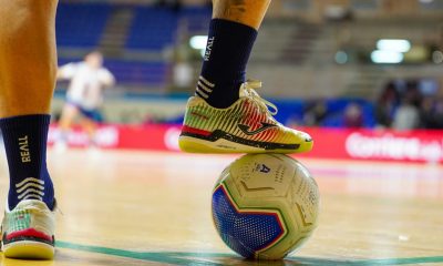
Walking and weight training alone are not enough for strong bones – football and handball may be more effective
Scheduling dental care during prenatal visits boosts oral health care during pregnancy
Enjoying the grilling at Mandaue City’s Matias BBQ
Study links coffee consumption to metabolic health and sex hormones
Dogs can distinguish between human fear and sadness
Manulife Investments Philippines leads UITF rankings across equity and fixed income funds
The look and feel of your neighborhood may affect your sleep
Pasalubong shopping in Davao via Aldevinco
Workout or nap? Either can help your sleepless brain, study finds

-
 NewsMakers3 weeks ago
Scheduling dental care during prenatal visits boosts oral health care during pregnancy
-
 Destinations3 weeks ago
Enjoying the grilling at Mandaue City’s Matias BBQ
-
 NewsMakers3 weeks ago
Study links coffee consumption to metabolic health and sex hormones
-
 Pet Care3 weeks ago
Dogs can distinguish between human fear and sadness
-
 Product Showcase3 weeks ago
Manulife Investments Philippines leads UITF rankings across equity and fixed income funds
-
 NewsMakers1 week ago
The look and feel of your neighborhood may affect your sleep
-
 Destinations4 days ago
Pasalubong shopping in Davao via Aldevinco
-
 NewsMakers1 week ago
Workout or nap? Either can help your sleepless brain, study finds