
NewsMakers
Scientists find evidence that novel coronavirus infects the mouth’s cells
“When infected saliva is swallowed or tiny particles of it are inhaled, we think it can potentially transmit SARS-CoV-2 further into our throats, our lungs, or even our guts.”


An international team of scientists has found evidence that SARS-CoV-2, the virus that causes COVID-19, infects cells in the mouth. While it’s well known that the upper airways and lungs are primary sites of SARS-CoV-2 infection, there are clues the virus can infect cells in other parts of the body, such as the digestive system, blood vessels, kidneys and, as this new study shows, the mouth.
The potential of the virus to infect multiple areas of the body might help explain the wide-ranging symptoms experienced by COVID-19 patients, including oral symptoms such as taste loss, dry mouth and blistering. Moreover, the findings point to the possibility that the mouth plays a role in transmitting SARS-CoV-2 to the lungs or digestive system via saliva laden with virus from infected oral cells. A better understanding of the mouth’s involvement could inform strategies to reduce viral transmission within and outside the body. The team was led by researchers at the National Institutes of Health and the University of North Carolina at Chapel Hill.
“Due to NIH’s all-hands-on-deck response to the pandemic, researchers at the National Institute of Dental and Craniofacial Research were able to quickly pivot and apply their expertise in oral biology and medicine to answering key questions about COVID-19,” said NIDCR Director Rena D’Souza, D.D.S., M.S., Ph.D. “The power of this approach is exemplified by the efforts of this scientific team, who identified a likely role for the mouth in SARS-CoV-2 infection and transmission, a finding that adds to knowledge critical for combatting this disease.”
The study, published online March, 25, 2021 in Nature Medicine, was led by Blake M. Warner, D.D.S., Ph.D., M.P.H., assistant clinical investigator and chief of NIDCR’s Salivary Disorders Unit, and Kevin M. Byrd, D.D.S., Ph.D., at the time an assistant professor in the Adams School of Dentistry at the University of North Carolina at Chapel Hill. Byrd is now an Anthony R. Volpe Research Scholar at the American Dental Association Science and Research Institute. Ni Huang, Ph.D., of the Wellcome Sanger Institute in Cambridge, U.K., and Paola Perez, Ph.D., of NIDCR, were co-first authors.
Researchers already know that the saliva of people with COVID-19 can contain high levels of SARS-CoV-2, and studies suggest that saliva testing is nearly as reliable as deep nasal swabbing for diagnosing COVID-19. What scientists don’t entirely know, however, is where SARS-CoV-2 in the saliva comes from. In people with COVID-19 who have respiratory symptoms, virus in saliva possibly comes in part from nasal drainage or sputum coughed up from the lungs. But according to Warner, that may not explain how the virus gets into the saliva of people who lack those respiratory symptoms.
“Based on data from our laboratories, we suspected at least some of the virus in saliva could be coming from infected tissues in the mouth itself,” Warner said.
To explore this possibility, the researchers surveyed oral tissues from healthy people to identify mouth regions susceptible to SARS-CoV-2 infection. Vulnerable cells contain RNA instructions for making “entry proteins” that the virus needs to get into cells. RNA for two key entry proteins–known as the ACE2 receptor and the TMPRSS2 enzyme–was found in certain cells of the salivary glands and tissues lining the oral cavity. In a small portion of salivary gland and gingival (gum) cells, RNA for both ACE2 and TMPRSS2 was expressed in the same cells. This indicated increased vulnerability because the virus is thought to need both entry proteins to gain access to cells.
“The expression levels of the entry factors are similar to those in regions known to be susceptible to SARS-CoV-2 infection, such as the tissue lining the nasal passages of the upper airway,” Warner said.
Once the researchers had confirmed that parts of the mouth are susceptible to SARS-CoV-2, they looked for evidence of infection in oral tissue samples from people with COVID-19. In samples collected at NIH from COVID-19 patients who had died, SARS-CoV-2 RNA was present in just over half of the salivary glands examined. In salivary gland tissue from one of the people who had died, as well as from a living person with acute COVID-19, the scientists detected specific sequences of viral RNA that indicated cells were actively making new copies of the virus–further bolstering the evidence for infection.
Once the team had found evidence of oral tissue infection, they wondered whether those tissues could be a source of the virus in saliva. This appeared to be the case. In people with mild or asymptomatic COVID-19, cells shed from the mouth into saliva were found to contain SARS-CoV-2 RNA, as well as RNA for the entry proteins.
To determine if virus in saliva is infectious, the researchers exposed saliva from eight people with asymptomatic COVID-19 to healthy cells grown in a dish. Saliva from two of the volunteers led to infection of the healthy cells, raising the possibility that even people without symptoms might transmit infectious SARS-CoV-2 to others through saliva.
Finally, to explore the relationship between oral symptoms and virus in saliva, the team collected saliva from a separate group of 35 NIH volunteers with mild or asymptomatic COVID-19. Of the 27 people who experienced symptoms, those with virus in their saliva were more likely to report loss of taste and smell, suggesting that oral infection might underlie oral symptoms of COVID-19.
Taken together, the researchers said, the study’s findings suggest that the mouth, via infected oral cells, plays a bigger role in SARS-CoV-2 infection than previously thought.
“When infected saliva is swallowed or tiny particles of it are inhaled, we think it can potentially transmit SARS-CoV-2 further into our throats, our lungs, or even our guts,” said Byrd.
More research will be needed to confirm the findings in a larger group of people and to determine the exact nature of the mouth’s involvement in SARS-CoV-2 infection and transmission within and outside the body.
“By revealing a potentially underappreciated role for the oral cavity in SARS-CoV-2 infection, our study could open up new investigative avenues leading to a better understanding of the course of infection and disease. Such information could also inform interventions to combat the virus and alleviate oral symptoms of COVID-19,” Warner said.




NewsMakers
Study links coffee consumption to metabolic health and sex hormones
Despite having a similar body mass index (BMI), individuals with higher coffee consumption had lower total and visceral fat and greater skeletal muscle mass than those who consumed less coffee.

Coffee is one of the world’s most widely consumed beverages, and previous research has linked its consumption to a lower risk of conditions such as type 2 diabetes and cardiovascular disease. However, the biological mechanisms behind these benefits remain unclear. A new Finnish study links habitual coffee consumption to healthier body composition and metabolic markers, while revealing distinct associations with sex hormones in men and women.
The study, conducted at the University of Oulu, analysed data from 2,264 participants aged 46 in the Northern Finland Birth Cohort 1966. Researchers examined how habitual coffee consumption was associated with circulating metabolites, cardiometabolic risk markers and sex hormones.
Despite having a similar body mass index (BMI), individuals with higher coffee consumption had lower total and visceral fat and greater skeletal muscle mass than those who consumed less coffee.
In both men and women, higher coffee consumption was correlated with lower circulating levels of branched-chain amino acids, biomarkers that have previously been linked to insulin resistance and an increased risk of type 2 diabetes when chronically elevated.
The strongest sex-specific associations were observed in men. Higher coffee consumption was linked to a more favourable glucose–insulin profile, higher concentrations of total and bioavailable testosterone, and increased levels of sex hormone-binding globulin (SHBG). At the same time, free testosterone and the free androgen index were modestly lower. In women, hormonal associations were more limited and were primarily characterised by higher SHBG and lower measures of free androgens.
“Coffee is consumed by millions of people every day, yet we still know surprisingly little about how it relates to our metabolism and hormones. What stood out in our findings was a distinct hormonal signature that didn’t disappear even after we took into account BMI and lifestyle factors, with several of these associations differing between men and women,” says Luca Verroest, lead author of the study and Doctoral Researcher at the University of Oulu.
The results suggest that hormonal pathways may partly explain the relationship between coffee consumption and metabolic health. However, as this was an observational study, the findings demonstrate associations rather than cause-and-effect relationships.
The study is particularly relevant in Finland, one of the world’s highest coffee-consuming countries, where annual consumption averages around 11.8 kilograms per person.
The researchers say the findings provide a foundation for future studies aimed at determining whether coffee itself drives these biological changes and identifying the compounds responsible. These questions are currently being investigated in animal models, with the long-term goal of progressing to human intervention studies. Further research will be needed before the findings could inform dietary recommendations.
The study, Associations of habitual coffee intake with testosterone and cardiometabolic markers: the Northern Finland Birth Cohort 1966 study, has been published in the European Journal of Nutrition.
NewsMakers
Scheduling dental care during prenatal visits boosts oral health care during pregnancy
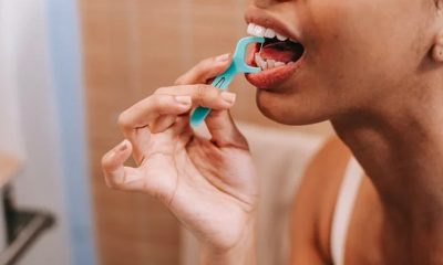
Scheduling a dental appointment during prenatal clinic visits—a simple, no-cost task that doesn’t require extra staff—increases the likelihood that patients later see a dentist during pregnancy.

Prenatal visits might include bloodwork, discussions of due dates and vitamins, and ultrasounds. What if these visits could also improve oral health care among pregnant patients?
New research shows that scheduling a dental appointment during prenatal clinic visits—a simple, no-cost task that doesn’t require extra staff—increases the likelihood that patients later see a dentist during pregnancy.
The study, published in the American Journal of Public Health, demonstrates the benefit of coordination among health providers and integrating oral health into prenatal care, especially for vulnerable patient populations.
Historically, dentists avoided treating women during pregnancy unless it was an emergency, or only provided care during the second trimester. This guidance changed once studies showed that routine and emergency dental care is safe during pregnancy—and current obstetric and dental guidelines stress that dental care can and should be provided during this critical period.
Moreover, pregnancy increases the risk of some oral health conditions, including gum inflammation and sometimes tooth decay, making routine dental care even more important during pregnancy. Some oral health issues may affect the fetus or even be transmissible to the child after birth.
Marginalized groups, including low-income individuals and those insured by Medicaid, are known to have higher rates of dental disease. Yet women enrolled in Medicaid are far less likely to see a dentist for a cleaning during pregnancy compared to women with private insurance.
“Many of those vulnerable to oral disease enter pregnancy with dental disease, and this may worsen during pregnancy,” said study author Stefanie Russell, associate professor at Rutgers School of Dental Medicine and an adjunct associate professor at NYU College of Dentistry, who led this research while at NYU.
One way to try and increase dental care among pregnant women is to reach them where they already are: their prenatal appointments. To bridge this gap, researchers and clinicians at NYU College of Dentistry teamed up with neighboring NYC Health + Hospitals/Bellevue, the oldest public hospital in the US, to create the New York University/Bellevue Prenatal Oral Health Program.
During weekly sessions at Bellevue’s prenatal clinic, pregnant patients receive a standardized screening to assess their oral health. Those with dental needs are referred to NYU College of Dentistry for follow-up care, educated about the importance and safety of dental care during pregnancy, and informed that Medicaid covers dental care in New York.
From 2018 to 2020, 420 prenatal patients with Medicaid coverage were referred to NYU Dentistry for follow-up dental care; the majority were Hispanic, and roughly half spoke Spanish as their first language. Referrals for dental care took two forms: immediately scheduled dental appointments made before the patient left the prenatal clinic or through a patient navigator. The patient navigator—a bilingual professional—educated patients regarding oral health, reminded patients of their upcoming dental appointments, and followed up after to identify any issues.
Nearly 44 percent of patients referred to NYU College of Dentistry followed through with a dental appointment. Both immediate appointment scheduling and the patient navigator increased the chances of a patient receiving dental care, but those who had appointments scheduled at their prenatal visit were 2.6 times more likely to see a dentist than those without appointments scheduled—even more likely than those who worked with the patient navigator.
“This suggests that immediately scheduled dental appointments can be an effective way to increase dental care utilization during pregnancy, without needing to hire extra staff,” said study author Shulamite Huang, assistant professor of epidemiology and health promotion at NYU College of Dentistry.
The authors conclude that integrating oral health screening in a prenatal clinic is feasible and increases engagement with dental care during pregnancy. They are continuing their research to measure whether this care improves oral health outcomes and to better understand how dental screening can be successfully integrated with prenatal care in other settings.
“A warm hand-off is key for addressing part of the barriers to dental care during pregnancy. Our team plans to further investigate and optimize interventions addressing both patient-, provider-, and healthcare system-level barriers to dental care during pregnancy,” said Huang.
Additional study authors include Richard Heyman and Mary Kang of NYU Dentistry; Belkys Saba, formerly of NYU Dentistry; Chloe Bird of the Tufts University School of Medicine; and Chengwu Yang of the Florida Atlantic University Schmidt College of Medicine.
NewsMakers
Why a UTI hurts — and why that might be a good thing
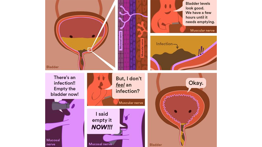
Australian researchers have discovered a previously overlooked group of bladder nerves that help detect urinary tract infections (UTIs) and trigger the body’s response to clear them, providing a potential new target for future bladder pain therapies.
Australian researchers have discovered a previously overlooked group of bladder nerves that help detect urinary tract infections (UTIs) and trigger the body’s response to clear them, providing a potential new target for future bladder pain therapies.
The study shows that bladder nerves located close to the lining of the bladder act as a frontline infection sensor, helping the body recognise UTIs and trigger responses that reduce the severity and spread of infection.
UTIs are among the most common bacterial infections worldwide, with more than 400 million cases reported every year. Nearly one in three women will experience UTIs before the age of 24, and many elderly people and those with bladder issues from spinal cord injuries can experience multiple UTIs in a single year.
Symptoms often include frequent urination, a sudden urge to urinate, pain during urination, and pelvic discomfort can be debilitating for some patients.
Flinders University’s Dr Luke Grundy says that while scientists have long understood how the bladder senses as it fills and triggers urination, the role of a specialised group of bladder nerves near the bladder lining has remained unclear.
“Most bladder nerves act like a fuel gauge, telling the brain when the bladder is filling up and needs emptying,” says Dr Grundy, Head of the NeuroUrology Research Group at Flinders University.
“The nerves we studied in this research are different. They sit close to the bladder lining and appear to act more like an early warning system, detecting infection and inflammation.
“They don’t just sense infection. They help coordinate the body’s response to it by triggering pain and urinary frequency, behaviours that appear to help clear bacteria from the bladder as part of the body’s defence system.”
Lead author and recently graduated PhD student Dr Cindy Tay says the discovery changes how these nerves are understood.
“These mucosal nerves have puzzled scientists for almost two decades because they stay quiet while the bladder fills and empties, which is the main job of the bladder,” says Dr Tay.
“What we’ve found is that they have a hidden job — acting as an early warning system that springs into action the moment infection takes hold.”
The research team developed a novel method to selectively study a specialised group of sensory nerves in the bladder lining of mice, revealing that while these nerves play little role in normal bladder function, they become highly responsive during a UTI and help detect and respond to infection.
“When the bladder is healthy, these nerves are relatively quiet, but during a urinary tract infection they become highly sensitive and respond to the presence of bacteria and inflammation,” says Dr Grundy.
“It may feel unpleasant, but urinating more frequently actually helps clear the infection by flushing out the harmful bacteria.”
The study also helps explain why people with conditions affecting nerve function may be more prone to recurrent or severe UTIs.
“If the nerves that detect infection aren’t working properly, the body may not respond as effectively,” says Dr Grundy.
Building on previous research, the new study reveals a deeper understanding of how UTIs affect bladder function and the nervous system, and could help develop new treatments that target these nerves to relieve UTI-related symptoms.
“Our findings provide new insight into how the bladder detects and responds to infection, helping explain the biological processes that drive the pain, urgency and discomfort commonly experienced during UTIs,” says Dr Grundy
Researchers say the next challenge is to develop therapies that ease the pain and urgency associated with UTIs while preserving the protective role these nerves play in fighting infection.
The paper, ‘Bladder mucosal afferents detect UTI and aid pathogen clearance,’ by Cindy Tay, Harman Sharma, Stewart Ramsay (University of Adelaide), Georgia Bourlotos, Sarah K Manning, Natalie E Stevens, Sophie J Miller, Geraint B Rogers, David J Lynn, Feargal J Ryan, Andrea M Harrington (University of Adelaide), Vladimir Zagorodnyuk, Steven L Taylor and Luke Grundy was published in Proceedings of the National Academy of Sciences (PNAS).

Study links coffee consumption to metabolic health and sex hormones

Manulife Investments Philippines leads UITF rankings across equity and fixed income funds

Scheduling dental care during prenatal visits boosts oral health care during pregnancy

Enjoying the grilling at Mandaue City’s Matias BBQ

Dogs can distinguish between human fear and sadness

Pet dog microbiota may protect young children from respiratory tract infections and reduce the need for antibiotic courses

Walking and weight training alone are not enough for strong bones – football and handball may be more effective

Keeping kitty indoors is not the cat-astrophe that owners fear

Why a UTI hurts — and why that might be a good thing

Pregnancy complications can signal heart disease risk years before traditional screening, study finds

Filipinos prioritize independence as legacy for longer lives

Coffee and heart health: How many cups of caffeinated coffee are safe to drink each day?

Going Chinese in Pulilan, Bulacan

Eating out in Cebu in one place via Pusô Village
Pregnancy complications can signal heart disease risk years before traditional screening, study finds
Why a UTI hurts — and why that might be a good thing

Posture can influence your mood and behavior, study suggests
Walking and weight training alone are not enough for strong bones – football and handball may be more effective
Pet dog microbiota may protect young children from respiratory tract infections and reduce the need for antibiotic courses
Dogs can distinguish between human fear and sadness

-
 NewsMakers3 weeks ago
Filipinos prioritize independence as legacy for longer lives
-
 NewsMakers4 weeks ago
Coffee and heart health: How many cups of caffeinated coffee are safe to drink each day?
-
 Destinations3 weeks ago
Going Chinese in Pulilan, Bulacan
-
 Destinations2 weeks ago
Eating out in Cebu in one place via Pusô Village
-
 NewsMakers2 weeks ago
Pregnancy complications can signal heart disease risk years before traditional screening, study finds
-
 NewsMakers2 weeks ago
Why a UTI hurts — and why that might be a good thing
-
 NewsMakers2 weeks ago
Posture can influence your mood and behavior, study suggests
-
 Wellness2 weeks ago
Walking and weight training alone are not enough for strong bones – football and handball may be more effective