
NewsMakers
Placentas from COVID-19-positive pregnant women show injury
The type of injury seen in the placentas shows abnormal blood flow between the mothers and their babies in utero, pointing to a new complication of COVID-19.


The placentas from 16 women who tested positive for COVID-19 while pregnant showed evidence of injury, according to pathological exams completed directly following birth, reports a new Northwestern Medicine study.
The type of injury seen in the placentas shows abnormal blood flow between the mothers and their babies in utero, pointing to a new complication of COVID-19. The findings, though early, could help inform how pregnant women should be clinically monitored during the pandemic.
The study was published today (May 22) in the journal American Journal of Clinical Pathology. It is the largest study to examine the health of placentas in women who tested positive for COVID-19.
“Most of these babies were delivered full-term after otherwise normal pregnancies, so you wouldn’t expect to find anything wrong with the placentas, but this virus appears to be inducing some injury in the placenta,” said senior author Dr. Jeffrey Goldstein, assistant professor of pathology at Northwestern University Feinberg School of Medicine and a Northwestern Medicine pathologist. “It doesn’t appear to be inducing negative outcomes in live-born infants, based on our limited data, but it does validate the idea that women with COVID should be monitored more closely.”
This increased monitoring might come in the form of non-stress tests, which examine how well the placenta is delivering oxygen, or growth ultrasounds, which measure if the baby is growing at a healthy rate, said co-author Dr. Emily Miller, assistant professor of obstetrics and gynecology at Feinberg and a Northwestern Medicine obstetrician.
“Not to paint a scary picture, but these findings worry me,” Miller said. “I don’t want to draw sweeping conclusions from a small study, but this preliminary glimpse into how COVID-19 might cause changes in the placenta carries some pretty significant implications for the health of a pregnancy. We must discuss whether we should change how we monitor pregnant women right now.”
Previous research has found that children who were in utero during the 1918-19 flu pandemic, which is often compared to the current COVID-19 pandemic, have lifelong lower incomes and higher rates of cardiovascular disease. Flu doesn’t cross the placenta, Goldstein said, so whatever is causing life-long problems in those people is most likely due to immune activity and injury to the placenta.
“Our study, and other studies like it, are trying to get on the ground floor for this exposure so we can think about what research questions we should be asking in these kids and what can or should we do now to mitigate these same types of outcomes,” Goldstein said.
Fifteen patients delivered live infants in the third trimester, however one patient had a miscarriage in the second trimester. “That patient was asymptomatic, so we don’t know whether the virus caused the miscarriage or it was unrelated,” Goldstein said, “We are aware of four other cases of miscarriage with COVID. The other reported patients had symptoms and three of four had severe inflammation in the placenta. I’d like to see more before drawing any conclusions.”
The placenta is the first organ to form in fetal development. It acts as the fetus’ lungs, gut, kidneys and liver, taking oxygen and nutrients from the mother’s blood stream and exchanging waste. The placenta also is responsible for many of the hormonal changes within the mother’s body. Examining a woman’s placenta allows a pathologist to follow a retroactive roadmap of a woman’s pregnancy to learn what happened to the baby in utero or what could happen to both the mother and the infant after birth.
“The placenta acts like a ventilator for the fetus, and if it gets damaged, there can be dire outcomes,” Miller said. “In this very limited study, these findings provide some signs that the ventilator might not work as well for as long as we’d like it to if the mother tests positive for SARS-CoV2.”
The placentas in these patients had two common abnormalities: insufficient blood flow from the mother to the fetus with abnormal blood vessels called maternal vascular malperfusion (MVM) and blood clots in the placenta, called intervillous thrombi.
In normal cases of MVM, the mother’s blood pressure is higher than normal. This condition is typically seen in women with preeclampsia or hypertension. Interestingly, only one of the 15 patients in this study had preeclampsia or hypertension.
“There is an emerging consensus that there are problems with coagulation and blood vessel injury in COVID-19 patients,” Goldstein said. “Our finding support that there might be something clot-forming about coronavirus, and it’s happening in the placenta.”
The 16 women in the study delivered their babies at Northwestern Medicine Prentice Women’s Hospital. All tested positive for COVID-19. Four patients came in with flu-like symptoms three to five weeks before delivery and tested positive for the virus. The remaining patients all tested positive when they came in to deliver. Five patients never developed symptoms, others were symptomatic at delivery.
Between 30 and 40 patients deliver at Prentice daily. The team began testing placentas of COVID-19-positive mothers in early April. Fourteen of the live-born infants in the study were born full term and with normal weights and Apgar scores. One live-born infant was premature.
“They were healthy, full-term, beautifully normal babies, but our findings indicate a lot of the blood flow was blocked off and many of the placentas were smaller than they should have been,” Miller said. “Placentas get built with an enormous amount of redundancy. Even with only half of it working, babies are often completely fine. Still, while most babies will be fine, there’s a risk that some pregnancies could be compromised.”
In February, before the pandemic was known to have reached Chicago, Goldstein assembled his research team.
“If you get the flu and you’re pregnant, we know nothing about what that looks like in your placenta, so I began thinking how we’d study this flu-like epidemic if it came through Chicago,” Goldstein said. “We started setting things up and then lo and behold, the epidemic came here, so we were ready.”
Other Northwestern co-authors include Elisheva D. Shanes, Leena B. Mithal and Hooman A. Azad.





NewsMakers
Scheduling dental care during prenatal visits boosts oral health care during pregnancy
Scheduling a dental appointment during prenatal clinic visits—a simple, no-cost task that doesn’t require extra staff—increases the likelihood that patients later see a dentist during pregnancy.

Prenatal visits might include bloodwork, discussions of due dates and vitamins, and ultrasounds. What if these visits could also improve oral health care among pregnant patients?
New research shows that scheduling a dental appointment during prenatal clinic visits—a simple, no-cost task that doesn’t require extra staff—increases the likelihood that patients later see a dentist during pregnancy.
The study, published in the American Journal of Public Health, demonstrates the benefit of coordination among health providers and integrating oral health into prenatal care, especially for vulnerable patient populations.
Historically, dentists avoided treating women during pregnancy unless it was an emergency, or only provided care during the second trimester. This guidance changed once studies showed that routine and emergency dental care is safe during pregnancy—and current obstetric and dental guidelines stress that dental care can and should be provided during this critical period.
Moreover, pregnancy increases the risk of some oral health conditions, including gum inflammation and sometimes tooth decay, making routine dental care even more important during pregnancy. Some oral health issues may affect the fetus or even be transmissible to the child after birth.
Marginalized groups, including low-income individuals and those insured by Medicaid, are known to have higher rates of dental disease. Yet women enrolled in Medicaid are far less likely to see a dentist for a cleaning during pregnancy compared to women with private insurance.
“Many of those vulnerable to oral disease enter pregnancy with dental disease, and this may worsen during pregnancy,” said study author Stefanie Russell, associate professor at Rutgers School of Dental Medicine and an adjunct associate professor at NYU College of Dentistry, who led this research while at NYU.
One way to try and increase dental care among pregnant women is to reach them where they already are: their prenatal appointments. To bridge this gap, researchers and clinicians at NYU College of Dentistry teamed up with neighboring NYC Health + Hospitals/Bellevue, the oldest public hospital in the US, to create the New York University/Bellevue Prenatal Oral Health Program.
During weekly sessions at Bellevue’s prenatal clinic, pregnant patients receive a standardized screening to assess their oral health. Those with dental needs are referred to NYU College of Dentistry for follow-up care, educated about the importance and safety of dental care during pregnancy, and informed that Medicaid covers dental care in New York.
From 2018 to 2020, 420 prenatal patients with Medicaid coverage were referred to NYU Dentistry for follow-up dental care; the majority were Hispanic, and roughly half spoke Spanish as their first language. Referrals for dental care took two forms: immediately scheduled dental appointments made before the patient left the prenatal clinic or through a patient navigator. The patient navigator—a bilingual professional—educated patients regarding oral health, reminded patients of their upcoming dental appointments, and followed up after to identify any issues.
Nearly 44 percent of patients referred to NYU College of Dentistry followed through with a dental appointment. Both immediate appointment scheduling and the patient navigator increased the chances of a patient receiving dental care, but those who had appointments scheduled at their prenatal visit were 2.6 times more likely to see a dentist than those without appointments scheduled—even more likely than those who worked with the patient navigator.
“This suggests that immediately scheduled dental appointments can be an effective way to increase dental care utilization during pregnancy, without needing to hire extra staff,” said study author Shulamite Huang, assistant professor of epidemiology and health promotion at NYU College of Dentistry.
The authors conclude that integrating oral health screening in a prenatal clinic is feasible and increases engagement with dental care during pregnancy. They are continuing their research to measure whether this care improves oral health outcomes and to better understand how dental screening can be successfully integrated with prenatal care in other settings.
“A warm hand-off is key for addressing part of the barriers to dental care during pregnancy. Our team plans to further investigate and optimize interventions addressing both patient-, provider-, and healthcare system-level barriers to dental care during pregnancy,” said Huang.
Additional study authors include Richard Heyman and Mary Kang of NYU Dentistry; Belkys Saba, formerly of NYU Dentistry; Chloe Bird of the Tufts University School of Medicine; and Chengwu Yang of the Florida Atlantic University Schmidt College of Medicine.
NewsMakers
Why a UTI hurts — and why that might be a good thing
Australian researchers have discovered a previously overlooked group of bladder nerves that help detect urinary tract infections (UTIs) and trigger the body’s response to clear them, providing a potential new target for future bladder pain therapies.
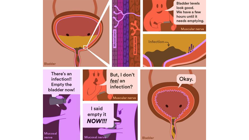
Australian researchers have discovered a previously overlooked group of bladder nerves that help detect urinary tract infections (UTIs) and trigger the body’s response to clear them, providing a potential new target for future bladder pain therapies.
The study shows that bladder nerves located close to the lining of the bladder act as a frontline infection sensor, helping the body recognise UTIs and trigger responses that reduce the severity and spread of infection.
UTIs are among the most common bacterial infections worldwide, with more than 400 million cases reported every year. Nearly one in three women will experience UTIs before the age of 24, and many elderly people and those with bladder issues from spinal cord injuries can experience multiple UTIs in a single year.
Symptoms often include frequent urination, a sudden urge to urinate, pain during urination, and pelvic discomfort can be debilitating for some patients.
Flinders University’s Dr Luke Grundy says that while scientists have long understood how the bladder senses as it fills and triggers urination, the role of a specialised group of bladder nerves near the bladder lining has remained unclear.
“Most bladder nerves act like a fuel gauge, telling the brain when the bladder is filling up and needs emptying,” says Dr Grundy, Head of the NeuroUrology Research Group at Flinders University.
“The nerves we studied in this research are different. They sit close to the bladder lining and appear to act more like an early warning system, detecting infection and inflammation.
“They don’t just sense infection. They help coordinate the body’s response to it by triggering pain and urinary frequency, behaviours that appear to help clear bacteria from the bladder as part of the body’s defence system.”
Lead author and recently graduated PhD student Dr Cindy Tay says the discovery changes how these nerves are understood.
“These mucosal nerves have puzzled scientists for almost two decades because they stay quiet while the bladder fills and empties, which is the main job of the bladder,” says Dr Tay.
“What we’ve found is that they have a hidden job — acting as an early warning system that springs into action the moment infection takes hold.”
The research team developed a novel method to selectively study a specialised group of sensory nerves in the bladder lining of mice, revealing that while these nerves play little role in normal bladder function, they become highly responsive during a UTI and help detect and respond to infection.
“When the bladder is healthy, these nerves are relatively quiet, but during a urinary tract infection they become highly sensitive and respond to the presence of bacteria and inflammation,” says Dr Grundy.
“It may feel unpleasant, but urinating more frequently actually helps clear the infection by flushing out the harmful bacteria.”
The study also helps explain why people with conditions affecting nerve function may be more prone to recurrent or severe UTIs.
“If the nerves that detect infection aren’t working properly, the body may not respond as effectively,” says Dr Grundy.
Building on previous research, the new study reveals a deeper understanding of how UTIs affect bladder function and the nervous system, and could help develop new treatments that target these nerves to relieve UTI-related symptoms.
“Our findings provide new insight into how the bladder detects and responds to infection, helping explain the biological processes that drive the pain, urgency and discomfort commonly experienced during UTIs,” says Dr Grundy
Researchers say the next challenge is to develop therapies that ease the pain and urgency associated with UTIs while preserving the protective role these nerves play in fighting infection.
The paper, ‘Bladder mucosal afferents detect UTI and aid pathogen clearance,’ by Cindy Tay, Harman Sharma, Stewart Ramsay (University of Adelaide), Georgia Bourlotos, Sarah K Manning, Natalie E Stevens, Sophie J Miller, Geraint B Rogers, David J Lynn, Feargal J Ryan, Andrea M Harrington (University of Adelaide), Vladimir Zagorodnyuk, Steven L Taylor and Luke Grundy was published in Proceedings of the National Academy of Sciences (PNAS).
NewsMakers
Pregnancy complications can signal heart disease risk years before traditional screening, study finds
Some women, particularly younger women often considered low risk, may face a higher risk of heart disease earlier than previously recognized.

McGill University researchers have developed a new tool to identify heart disease risk in women earlier in life.
Findings from a study published in JACC: Advances highlight gaps in existing approaches.
“Heart disease is the leading cause of death in women, yet existing risk tools were developed in older populations and ignore factors unique to women,” said senior author Robert Platt, Professor in the Department of Epidemiology, Biostatistics, and Occupational Health and Director of the School of Population and Global Health.
While pregnancy complications are known to be linked to future heart risk, there has been no way to identify which younger women are most at risk, he added.
Detecting risk earlier
Using health data from more than 260,000 women in the UK aged 15 to 45 who had given birth, researchers developed and validated a prediction model to estimate future heart disease risk. Participants were followed for nearly four years after delivery.
The model identified several factors – not included in existing tools – that can help predict risk, including hypertensive disorders of pregnancy, gestational diabetes, preterm birth, PCOS, depression, thyroid disorders, oral contraceptive use and social deprivation.
The findings suggest some women, particularly younger women often considered low risk, may face a higher risk of heart disease earlier than previously recognized.
“Millions of women who give birth each year are never considered candidates for cardiovascular risk assessment simply because of their age,” said co-author Kristian Filion, Professor in the Departments of Medicine and of Epidemiology, Biostatistics, and Occupational Health.
If integrated into routine postpartum care, this tool could enable earlier monitoring, lifestyle counselling or referral to a specialist, potentially helping prevent a heart attack or stroke later in life, he added.
The next step is to validate the model in Canada and the United States. In the longer term, the goal is to integrate a practical calculator into electronic health records so higher-risk patients can be identified earlier.
About the study
“Development and Validation of a Prediction Model for Cardiovascular Risk in Reproductive-Aged Women” by Sonia Grandi, Kristian Filion, Jennifer Hutcheon, Graeme Smith, and Robert Platt was published in JACC: Advances. The study was supported by the Canadian Institutes of Health Research.

Manulife Investments Philippines leads UITF rankings across equity and fixed income funds

Scheduling dental care during prenatal visits boosts oral health care during pregnancy

Enjoying the grilling at Mandaue City’s Matias BBQ

Dogs can distinguish between human fear and sadness

Pet dog microbiota may protect young children from respiratory tract infections and reduce the need for antibiotic courses

Walking and weight training alone are not enough for strong bones – football and handball may be more effective

Keeping kitty indoors is not the cat-astrophe that owners fear

Why a UTI hurts — and why that might be a good thing

Pregnancy complications can signal heart disease risk years before traditional screening, study finds

Posture can influence your mood and behavior, study suggests

Filipinos prioritize independence as legacy for longer lives

Coffee and heart health: How many cups of caffeinated coffee are safe to drink each day?

Going Chinese in Pulilan, Bulacan

Eating out in Cebu in one place via Pusô Village
Pregnancy complications can signal heart disease risk years before traditional screening, study finds
Posture can influence your mood and behavior, study suggests
Why a UTI hurts — and why that might be a good thing
Walking and weight training alone are not enough for strong bones – football and handball may be more effective
Pet dog microbiota may protect young children from respiratory tract infections and reduce the need for antibiotic courses
Keeping kitty indoors is not the cat-astrophe that owners fear

-
 NewsMakers3 weeks ago
Filipinos prioritize independence as legacy for longer lives
-
 NewsMakers3 weeks ago
Coffee and heart health: How many cups of caffeinated coffee are safe to drink each day?
-
 Destinations3 weeks ago
Going Chinese in Pulilan, Bulacan
-
 Destinations2 weeks ago
Eating out in Cebu in one place via Pusô Village
-
 NewsMakers2 weeks ago
Pregnancy complications can signal heart disease risk years before traditional screening, study finds
-
 NewsMakers2 weeks ago
Posture can influence your mood and behavior, study suggests
-
 NewsMakers2 weeks ago
Why a UTI hurts — and why that might be a good thing
-
 Wellness2 weeks ago
Walking and weight training alone are not enough for strong bones – football and handball may be more effective